
Most cases of BE are diagnosed using a non-invasive, high-resolution chest CT scan without contrast. While a standard chest X-ray may show some lung abnormalities, CT scans offer greater precision in identifying BE. The CT scan shows exactly which parts of the lungs are affected and how significantly the disease has progressed, both of which help determine the best treatments. CT scans also reveal distinctive patterns or appearances of BE, such as “tree-in-bud,” which describes the appearance of a small airway surrounded by tiny bud-like nodules resembling a tree branch. This pattern reflects inflammation in the small airways, usually due to infection and mucus impaction, and highlights the need for airway clearance – both to test for a bacterial infection and to remove the stagnant secretions.
Nodular bronchiectasis, characterized by small, clustered airway nodules, and cavitary bronchiectasis, marked by thick-walled air-filled spaces, are more commonly associated with recurrent respiratory infections, particularly those caused by nontuberculous mycobacteria (NTM).
Other descriptions of BE on a CT scan include the shape, often referred to as morphology:
1. Cylindrical: milder form of BE where the bronchus is the correct shape
but does not taper properly
2. Varicoid: widened segments of the bronchus bulge in some areas, narrow
in others, causing an irregular shape. This form of BE tends to have a
higher secretion burden, meaning heavier mucus buildup in the airways.
3. Cystic: a severe form of BE where the airway is very dilated into a cyst-
like form. Patients with cystic BE tend to have high secretion burden.
A CT scan report may also refer to the extent or distribution of the BE:
1. Diffuse: BE affects most or all lobes of the lung
2. Localised: BE affects one or two lobes, or is localized to one lung
3. Focal: common in post-infectious BE, the bronchiectasis affects only one or two smaller areas of a lobe
Your provider will also order respiratory cultures, pulmonary function tests, and a basic panel of blood tests aimed at finding the cause of your
bronchiectasis. At a minimum, the workup should include:
• Immunoglobulin levels (to look for immune deficiency)
• IgE and ABPA testing (to look for allergic-related bronchiectasis caused
by Aspergillus)
• Sweat chloride test and/or genetic testing for cystic fibrosis
• Testing for alpha-1 antitrypsin deficiency
• Consideration of genetic testing for primary ciliary dyskinesia (PCD)
• Autoimmune blood tests (for rheumatoid arthritis, scleroderma, and
other systemic diseases)
• Sputum cultures to identify organisms
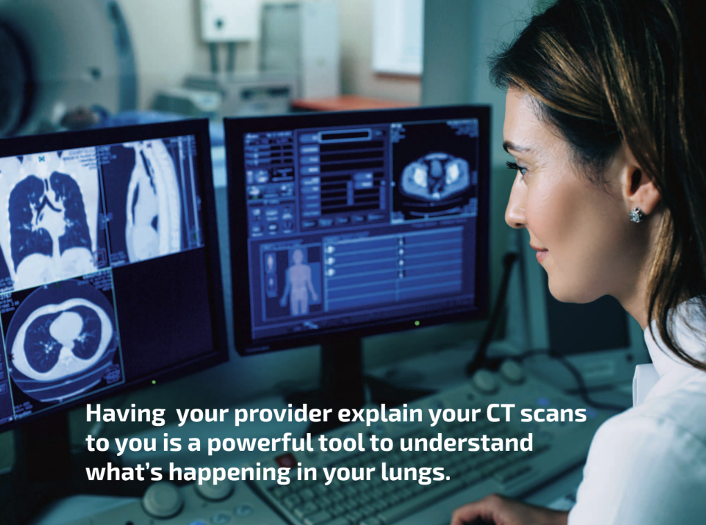
The frequency of CT scans depends on your disease severity and the recommendations of your provider. For some people with BE, a scan every three to five years may be sufficient. For others with more active disease, a CT scan every one to two years – or in some cases, every six months – may be needed. Your care team will help you determine how often they are needed. Consider asking your provider to show you your CT scans and explain them to you; understanding what is visually happening in your lungs is a powerful tool.