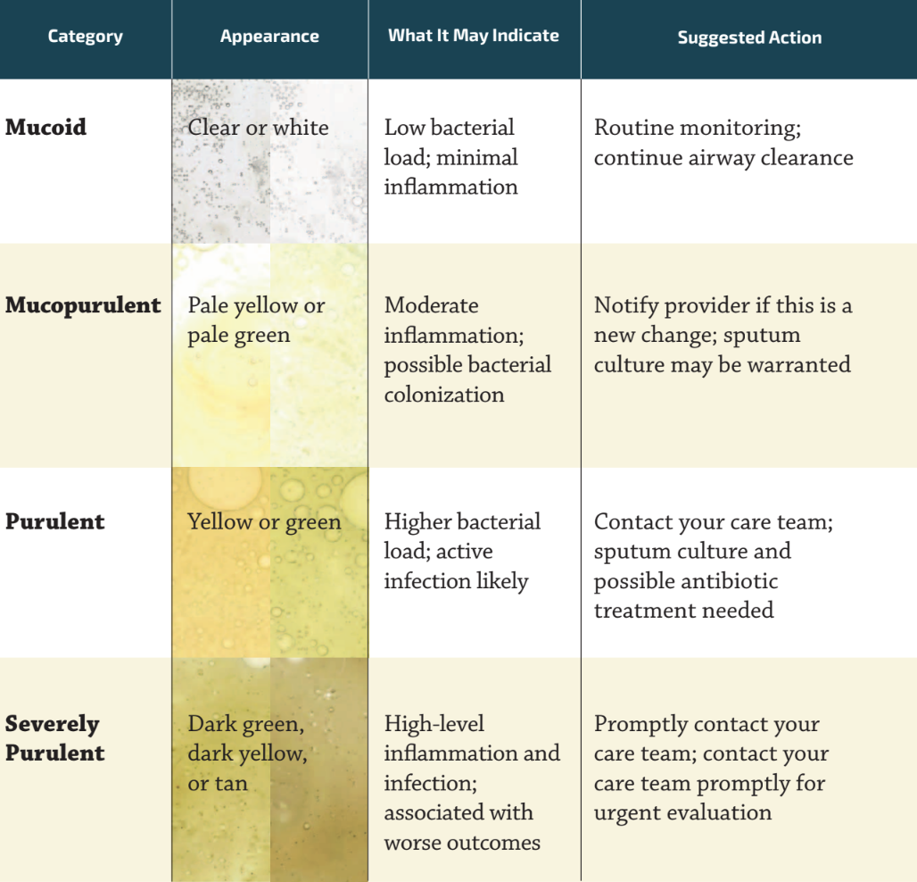
Sputum (Mucus) and What It Can Tell You One of the most practical things you can do to monitor your lung health is to pay attention to the color of your sputum (the mucus you cough up from your lungs). Sputum color is a simple, non-invasive indicator of what may be happening in your airways. In general, the greener or darker the sputum, the more inflammation and infection is present. A validated four-category sputum color chart (the Murray Sputum Color Chart) is used by clinicians and researchers to assess disease severity:
Murray Sputum Color Chart
The green color associated with more purulent sputum results from the accumulation of myeloperoxidase, an enzyme released by neutrophils
(immune cells) as they fight bacteria. More purulent sputum therefore reflects a higher degree of neutrophilic inflammation. Research has shown that sputum color reliably predicts bacterial colonization and the level of airway inflammation, and confirmed that sputum color is a meaningful marker of disease severity and future risk of exacerbations, hospitalization, and mortality.
Sputum color alone cannot identify which specific organism is present in your lungs, but it can provide clues about the level of inflammation. A formal sputum culture is needed to identify bacteria and guide treatment decisions. Track changes in your sputum color over time and report them to your care team. A shift from clear to yellow-green, especially alongside increased cough, fatigue, or shortness of breath, may signal an exacerbation.
Some colors are not captured on the standard chart and deserve special mention. Brown or rust-colored sputum can reflect old dried blood in the airways. Pink or frothy sputum can occasionally be seen in people with heart-related fluid accumulation. Black sputum may rarely be seen in heavy smokers or those with certain fungal infections. Any of these unusual colors should be reported to your provider.